Bahnbrechende Erkenntnisse der modernen Neurobiologie: Wie das menschliche Gehirn tatsächlich arbeitet und was die Forschung heute weiß

| Diets Premium Experte Mia Hoffmann | Min. Lesezeit 5 min | Wissenschaftlich geprüft durch klinische Ernährungsberater 23.10.2026 |
Audio-Sprecher
•Artikelinhalt live anhören
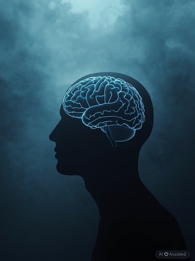
Mit rund 86 Milliarden Neuronen, ebenso vielen Gliazellen und über 100 Billionen synaptischen Verbindungen ist das menschliche Gehirn das komplexeste bekannte Organ. Obwohl es mit 1,3 bis 1,4 kg nur etwa 2 % des Körpergewichts ausmacht, beansprucht es rund 20 % des gesamten Ruheenergie- und Sauerstoffbedarfs. Dank moderner Bildgebung (fMRT, PET) und Optogenetik konnte die Hirnforschung hartnäckige Alltagsmythen widerlegen, das glymphatische Reinigungssystem im Schlaf entschlüsseln und völlig neue Therapieansätze gegen neurodegenerative Erkrankungen entwickeln.
Möchten Sie diese Richtlinien in Ihrer täglichen Ernährung umsetzen?
Unser intelligenter Diät-Generator erstellt in 3 Minuten einen maßgeschneiderten Speiseplan basierend auf diesen Empfehlungen.
Debunking the 10% Brain Myth: How Much of Our Neural Capacity Do We Actually Use?
One of the most pervasive neuromyths claims that humans utilize only 10% of their brain capacity. High-resolution functional magnetic resonance imaging (fMRI) and positron emission tomography (PET) have definitively refuted this misconception. Even during quiet rest, the brain’s intrinsic Default Mode Network (DMN) maintains widespread metabolic and electrical activity across cortical and subcortical regions, and over a 24-hour cycle, virtually 100% of the brain’s neural architecture is functionally engaged.
From an evolutionary bioenergetics standpoint, maintaining an organ that consumes 20% of the body’s total glucose and oxygen supply (350–450 kcal/day) would be biologically untenable if 90% of its tissue were dormant. Clinically, strokes or focal lesions affecting even a few millimeters of cerebral tissue consistently produce specific neurological deficits—confirming that no brain region lies idle.
Impact of Exogenous Cannabinoids (THC) on Adolescent Neuroarchitecture and Synaptic Pruning
While purified pharmaceutical cannabinoids (such as cannabidiol, CBD, in refractory pediatric epilepsies) hold validated medical indications, structural MRI investigations conducted by researchers at Northwestern and Harvard Universities demonstrated that regular recreational exposure to Δ9-tetrahydrocannabinol (THC) induces measurable morphological alterations in the nucleus accumbens, amygdala, and hippocampus.
Human cerebral maturation—specifically synaptic pruning and prefrontal cortical myelination—continues until approximately age 25, guided by endogenous cannabinoid signaling (anandamide and 2-AG acting on CB1 receptors). Chronic adolescent exposure to high-potency exogenous THC overstimulates CB1 receptors, disrupts prefrontal-limbic connectivity, impairs working memory and executive motivation, and significantly elevates the risk of psychosis in genetically vulnerable individuals.
Sleep Deprivation, the Glymphatic Clearance System, and Fatal Familial Insomnia (FFI)
The discovery of the brain’s glymphatic system by Dr. Maiken Nedergaard and colleagues fundamentally transformed our understanding of why sleep is biologically non-negotiable. During deep slow-wave NREM sleep, astrocytic glial cells shrink, expanding the cerebral interstitial space by roughly 60%. Driven by astrocytic aquaporin-4 (AQP4) water channels, cerebrospinal fluid flushes rapidly through brain parenchyma, clearing neurotoxic metabolic waste accumulated during wakefulness—most notably amyloid-beta (Aβ) peptides and hyperphosphorylated tau.
The catastrophic consequences of complete sleep loss are starkly illustrated by Fatal Familial Insomnia (FFI), a rare autosomal-dominant prion disease caused by a mutation in the PRNP gene. Misfolded prion proteins selectively destroy the mediodorsal and anterior nuclei of the thalamus, abolishing sleep spindles, triggering relentless sympathetic hyperactivity, and culminating in rapid neurodegeneration.
Optogenetics and Memory Engrams – How Neural Circuits Encode and Reconstruct Memories
For decades, neuroscientists sought to identify the physical substrate of a specific memory—the engram. The breakthrough arrived with the development of optogenetics, a technique that introduces light-sensitive microbial opsin ion channels (such as channelrhodopsin-2) into targeted neuronal populations, enabling millisecond-precision activation or silencing of specific neural circuits via fiber-optic laser pulses.
Nobel laureate Dr. Susumu Tonegawa and his team at MIT demonstrated in animal models that memories are encoded within discrete ensembles of hippocampal dentate gyrus and amygdala neurons. Crucially, human memory is not a static recording but a reconstructive biochemical process requiring protein synthesis during memory reconsolidation—a mechanism now leveraged in clinical psychotherapy to attenuate traumatic fear memories in post-traumatic stress disorder (PTSD).
The Blood-Brain Barrier and Novel Therapeutic Delivery in Neurodegenerative Diseases
A primary bottleneck in treating Alzheimer’s disease, Parkinson’s disease, and amyotrophic lateral sclerosis (ALS) is the blood-brain barrier (BBB)—a highly selective neurovascular unit formed by tight-junction endothelial cells, pericytes, and astrocytic endfeet. Historically, over 98% of small-molecule drugs and nearly 100% of large biologics (monoclonal antibodies, neurotrophic factors) failed to cross the BBB in therapeutic concentrations.
Cutting-edge neurotherapeutics now overcome this barrier via two translational strategies: receptor-mediated transcytosis („molecular Trojan horses”), which fuse therapeutic antibodies to transferrin or insulin receptor ligands to ferry cargo across endothelial cells, and MRI-guided focused ultrasound (FUS) combined with intravenous microbubbles, which transiently and non-invasively opens the BBB in targeted brain nuclei.
Circulating Plasma Factors, Adult Hippocampal Neurogenesis, and Nutritional Neuroprotection
Landmark studies from Harvard and Stanford Universities demonstrated that systemic vascular health and circulating plasma factors directly govern adult hippocampal neurogenesis and synaptic plasticity. Researchers identified specific blood-borne proteins—including tissue inhibitor of metalloproteinases-2 (TIMP2), exercise-induced myokine irisin, and brain-derived neurotrophic factor (BDNF)—that rejuvenate cerebral microvasculature and promote neural stem cell proliferation.
From a clinical preventive perspective, individuals can potently upregulate endogenous BDNF and preserve cerebrovascular integrity through three daily pillars: regular aerobic and resistance exercise, adherence to the neuroprotective MIND dietary pattern (supplying neuronal membrane DHA from marine fish, anthocyanins from berries, folate and lutein from leafy greens, extra-virgin olive oil, and walnuts), and prioritizing restorative slow-wave sleep.
Tags

Mia Hoffmann
✓Diets Premium Experte
Experte im Team von Diets Premium. Spezialisiert auf Ernährungspläne, Kraftsport und Trainingspläne.
Profil ansehen →