Hiatal hernia – symptoms, diagnosis, and diet for esophageal tumor hernia
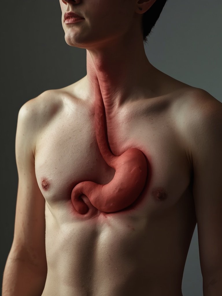
Table of Contents 4
Medical & Editorial Disclaimer (§3 Terms): This article is for informational and educational purposes only, does not constitute medical advice, diagnosis, or treatment, and is used at your own risk. Consult a qualified physician for medical conditions.
The comprehensive chapters outline the clinical spectrum and physiological consequences of this condition. Core symptoms range from persistent pyrosis and acid regurgitation to swallowing difficulties, morning hoarseness, and chest discomfort that can simulate non-cardiac chest pain. The underlying mechanism revolves around increased intra-abdominal pressure and mechanical instability of the esophageal junction. Management strategies balance conservative lifestyle changes, acid-suppressive pharmacotherapy, and surgical options for severe cases.
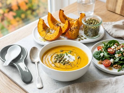
Practical guidance emphasizes anti-reflux dietary strategies to maintain digestive health. Consuming smaller, frequent meals throughout the day prevents gastric distension and reduces pressure on the lower esophageal sphincter. Restricting high-fat dishes, fried items, and aggressive spices aids in minimizing gastric acid secretion and esophageal mucosal irritation. Additionally, elevating the head during sleep, avoiding restrictive clothing around the abdomen, and allowing sufficient time between the final meal and bedtime leverage gravity to protect against nocturnal acid reflux.
A hiatal hernia, colloquially referred to as gastric displacement into the chest cavity, occurs when the proximal portion of the stomach protrudes through the diaphragmatic esophageal hiatus into the mediastinum. The diaphragm serves as the primary respiratory barrier separating the abdominal and thoracic compartments, featuring a small hiatus through which the esophagus normally reaches the gastric cardia. When this muscular opening widens and supporting phrenoesophageal ligaments weaken, the lower esophageal sphincter is compromised, leading to severe gastroesophageal reflux and requiring structured nutritional and medical management.
Anatomy, Etiology, and Types of Hiatal Hernia
Clinical classification distinguishes two primary forms of hiatal hernia: sliding hernias (accounting for more than 90% of diagnoses) and paraesophageal hernias. In sliding hiatal hernias, the gastroesophageal junction and the gastric cardia slide upward into the posterior mediastinum, often dynamically shifting with changes in intra-abdominal pressure and body posture. In paraesophageal hernias, the gastroesophageal junction remains anatomically situated below the diaphragm, but the gastric fundus rolls alongside the esophagus into the thorax, predisposing patients to volvulus, incarceration, and mucosal ischemia. Contributing factors include age-related connective tissue loss, visceral obesity, chronic coughing, straining, and elevated abdominal pressure.
Clinical Manifestations: From Heartburn to Pseudo-Anginal Chest Pain
The hallmark manifestations of a hiatal hernia stem from gastroesophageal reflux: persistent pyrosis (retrosternal burning), regurgitation of acidic stomach fluid, and postprandial discomfort exacerbated by bending forward or recumbent positioning. Patients frequently report dysphagia, odynophagia, morning hoarseness due to laryngeal acid exposure, and a nocturnal dry cough. Crucially, large hernias can provoke severe substernal pressure and radiation to the jaw or left shoulder that closely mimics acute coronary syndrome or myocardial infarction. Ruling out cardiac emergencies via urgent electrocardiography is mandatory before pursuing confirmatory endoscopy and esophageal manometry.
Nutritional Management and Anti-Reflux Dietary Protocols
Nutritional intervention represents the cornerstone of conservative hiatal hernia and GERD management. The diet must prioritize small, frequent meals (5–6 portions daily) eaten at regular intervals to prevent gastric distension and subsequent transient lower esophageal sphincter relaxations. The final meal of the day must occur at least 3 hours prior to sleep. Dietary triggers that relax sphincter tone or irritate esophageal mucosa must be restricted: deep-fried dishes, high-fat meats, fiery spices, citrus fruits, tomato paste, raw onions, garlic, peppermint, chocolate, caffeine, carbonated beverages, and alcohol. Cooking techniques should emphasize steaming, poaching, and stewing, with abundant incorporation of oatmeal, millet, lean poultry, non-acidic cooked vegetables, and soothing flaxseed mucilage.
Lifestyle Modifications and Surgical Indications: Fundoplication Techniques
Complementary lifestyle adjustments are indispensable: elevating the head of the bed by 15–20 cm (enlisting gravity to prevent nocturnal acid aspiration), avoiding tight belts and restrictive garments that raise intra-abdominal pressure, and refraining from recumbency or bending after eating. Progressive weight reduction in overweight patients provides dramatic, lasting symptomatic relief. Pharmacological management utilizes proton pump inhibitors (PPIs) and mucosal protectants. When conservative therapies fail, or in cases of large paraesophageal hernias harboring risks of strangulation or ulceration, laparoscopic surgical repair (such as Nissen or Toupet fundoplication combined with hiatoplasty) provides an effective anatomical cure.
Want to apply these guidelines in your daily diet?
Our smart diet generator creates a tailored meal plan in 3 minutes based on the recommendations in this article.
Tags

mgr Literatura: Chmiel K., Niśkiewicz I., Krela-Kaźmierczak I., Łykowska-Szuber L., Linke K. 2009. Pozaprzełykowe objawy refluksu żołądkowo-przełykowego. Nowiny Lekarskie, 78, 3-4, 216-221. Jarosz M. (red.) 2010. Praktyczny Podręcznik Dietetyki. Instytut Żywności i Żywienia, Warszawa. Pietrzak A. Refluks żołądkowo – przełykowy. 2016. Klinika Gastroenterologii, Hepatologii i Onkologii Klinicznej CMKP, 4-11, esoxxone.pl/data/2016/09/ESSOX-refluks-poradnik.pdf. Szelachowski P., Strutyńska-Karpińska M., Grabowski K., Taboła R., Pelczar P. 2006. Olbrzymia przepuklina rozworu przełykowego – opis przypadku. Advances in Clinical and Experimental Medicine, 15, 2, 389-391. Włodarek D., Lange E., Kozłowska L., Głąbska D. 2014. Dietoterapia. Wydawnictwo Lekarskie PZWL, Warszawa.
Diets Premium Expert
Diets Premium team expert. Specializes in nutrition plans, strength sports, and training plans.